array(2) { ["lab"]=> string(3) "793" ["publication"]=> string(4) "5117" } Mass spectrometry imaging of the in situ drug release from nanocarriers - 陈素明课题组(质谱研究组) | LabXing

陈素明课题组(质谱研究组)

简介 化学与生物质谱

分享到

Mass spectrometry imaging of the in situ drug release from nanocarriers

2018
期刊 Science Advances
下载全文
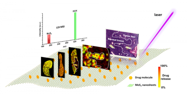
It is crucial but of a great challenge to study in vivo and in situ drug release of nanocarriers when developing a nanomaterial-based drug delivery platform. We developed a new label-free laser desorption/ionization mass spectrometry (MS) imaging strategy that enabled visualization and quantification of the in situ drug release in tissues by monitoring intrinsic MS signal intensity ratio of loaded drug over the nanocarriers. The proof of concept was demonstrated by investigating the doxorubicin (DOX)/polyethylene glycol–MoS2 nanosheets drug delivery system in tumor mouse models. The results revealed a tissue-dependent release behavior of DOX during circulation with the highest dissociation in tumor and lowest dissociation in liver tissues. The drug-loaded MoS2 nanocarriers are predominantly distributed in lung, spleen, and liver tissues, whereas the accumulation in the tumor was unexpectedly lower than in normal tissues. This new strategy could also be extended to other drug-carrier systems, such as carbon nanotubes and black phosphorus nanosheets, and opened a new path to evaluate the drug release of nanocarriers in the suborgan level.

  • 卷 4
  • 期 10
  • 页码 eaat9039
  • American Association for the Advancement of Science (AAAS)
  • ISSN: 2375-2548
  • DOI: 10.1126/sciadv.aat9039